
Why do limbs become deformed?
The limbs of dogs and cats, like the legs of people, are meant to be relatively straight. The mechanism by which puppies and kittens limbs develop and grow is very complex. Occasionally these processes go wrong and deformities result. There are zones at the ends of the bones in immature animals that are responsible for increasing bone length in an even (symmetrical) manner. These are called the growth plates (or ‘physes’).
Limb deformities may either be present at birth (congenital) or become apparent during growth (developmental). There are a number of possible causes of limb deformities, the two most common being a genetic disorder and injury (trauma). If a growth plate is injured, for example due to a fall, in a growing animal it may stop growing and cause the bone to become shortened and bent.
What are the most common types of growth deformity?
The fore limbs are more commonly affected than the hind limbs. This is partly because in the forearm (antebrachium) there are two bones that grow alongside each other (the radius and ulna). If one of these bones grows faster than the other, the limb will develop abnormally and become bent or twisted. Typically the paw will deviate outwards.
How are growth deformities investigated?
X-ray (radiography) is the principle method of investigating growth deformities. It enables assessment of the location of the deformity and also the direction and magnitude of the deformity. Computed tomography (CT) scanning provides cross-sectional images of the limb in all planes and multiple slices can be reformatted to provide a 3-dimensional image. This advanced imaging modality can provide important additional information compared to conventional X-rays. It is particularly useful for detecting abnormalities affecting joints.
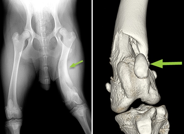
X-ray and CT scan of a Labradoodle showing deformity (bending) of the femur (thigh bone) (small arrow). The 3-dimensional CT scan enables the structure of the stifle (knee joint) to be assessed. In this dog the patella (knee cap) is displaced from the groove in the femur (large arrow).
How are growth deformities managed?
Many dogs and cats with mild growth deformities can be managed non-surgically. The deformity may be primarily cosmetic rather than causing pain and lameness. In these cases the appearance of the limb is not justification for surgery. In general, the smaller and more sedentary the patient, the greater should be the consideration for conservative management. In adult animals, corrective surgery is elective and thus can be considered at any time if the response to conservative management is unsatisfactory.
Conservative management may necessitate exercise restriction, such as avoidance of agility-type activities or jumping down from heights. This is especially important in immature dogs to reduce the possibility of further growth plate injury. Overweight animals should be placed on a diet. In dogs with remaining growth potential, the rate of growth should be evaluated and reduced if necessary by limiting food intake and considering changing feeding from a growth to an adult formula diet. Any dietary imbalances should be corrected.
Principles of surgery in immature dogs
The type of surgery will vary according to the age of the dog (since this influences the remaining growth potential), the bone or bones affected, the possibility of joint involvement, and the severity of the deformity. Certain important principles apply in all immature dogs with growth deformities:
- the problem should be assessed as early as possible
- the problem should (generally) be treated as soon as possible to prevent progressive deformity
- wherever possible the potential for continued growth should not be further compromised
The most common surgical procedures performed in immature dogs with growth deformities are explained below and comprise ostectomy (where there are paired bones), corrective osteotomy and limb lengthening.
Ostectomy of a paired bone
Retardation of growth of one of two paired bones may result in deformity and shortening of the remaining bone. Joint involvement is also possible. Removal of a section of the primarily affected bone (ostectomy) may be helpful in limiting further deformity in these cases, especially when there is significant remaining growth potential. Ostectomy of a paired bone is most commonly indicated in dogs less than six months of age. In a few cases it may be necessary to repeat the procedure if the ostectomy has healed and there is significant remaining growth potential.
Corrective osteotomy
Immature dogs with severe growth deformities often require early corrective osteotomy surgery in order to limit progressive deformity. A potential disadvantage of performing a corrective surgery on an immature patient is the possibility of further growth deformity, necessitating a second corrective procedure. The age (remaining growth potential) and size of the patient are important considerations.
Limb lengthening
Dogs can tolerate significant shortening of a bone without developing a significant gait abnormality. This is due to a) their ability to extend the joints in the affected limb, and b) compensatory overgrowth of another long bone in the affected limb. Limb lengthening, if required, is most readily achieved using a circular external skeletal fixator with tensioned wires that secure the two bone segments which need to be pulled away from each other (distracted). Specialist training is necessary for surgeons performing limb lengthening procedures, in order to minimise the potential for significant complications.
Principles of surgery in mature dogs
In mature dogs with growth deformities that require corrective surgery, the key considerations are:
-
-
- where should the corrective osteotomy (cutting of the bone) be performed?
- what degree of correction is required?
- how should the osteotomy (cut bone) be stabilised?
- is lengthening of the limb indicated?
-
The osteotomy (cutting of the bone) should be performed at the area of greatest deformity. The degree of correction is assessed both clinically and on X-rays (radiographically).
Osteotomy fixation
The most common methods of stabilising osteotomies (cut bones) are i) internal fixation with a bone plate and ii) external fixation with a fixator. Bone plates have certain mechanical advantages, are associated a low complication rate and rarely need to be removed. Disadvantages of bone plates include a) the inability to adjust limb alignment postoperatively and b) bone lengthening is not possible. External fixators have advantages of a) enabling limb lengthening and b) the ability to adjust limb alignment postoperatively. However, disadvantages include the need for pin tract maintenance and the necessity to remove the fixator.
Postoperative care and prognosis
Restriction of exercise following surgery is necessary until there is radiographic evidence of bone healing (usually six to eight weeks). Active and passive physiotherapy techniques are often beneficial.
The outlook or prognosis in dogs with growth deformities is dependant on a number of factors including the age of the dog, the degree of deformity/limb shortening, and the extent of joint involvement. The latter invariably results in osteoarthritis that may require ongoing medical management or occasionally necessitate salvage surgery, such as joint replacement or arthrodesis (fusion of the joint). In cases without joint involvement the prognosis is generally good.
Conclusion
In those dogs and cats where limb deformity surgery is indicated, the vast majority of patients benefit from the surgery. We will be pleased to give as much help and support as possible if you decide to give your pet the opportunity of limb deformity surgery.
Case study 1
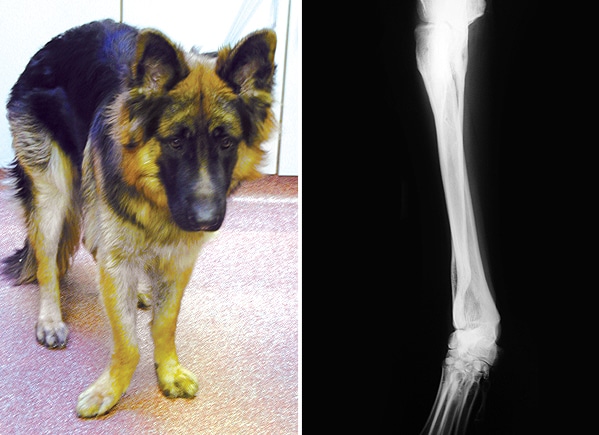
Photograph and X-ray of a German Shepherd Dog showing a deformed antebrachium (forearm). The right paw deviates outwards.
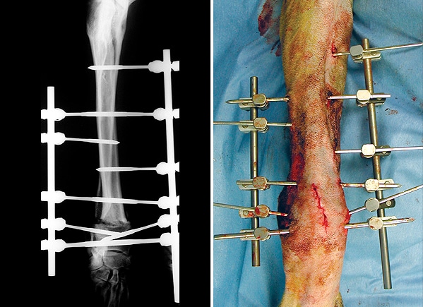
X-ray and photograph following surgery showing the straightened limb. The cut bones have been stabilised with pins placed through the skin and connected to bars on the inside and outside of the limb (known as an external skeletal fixator).
Case study 2
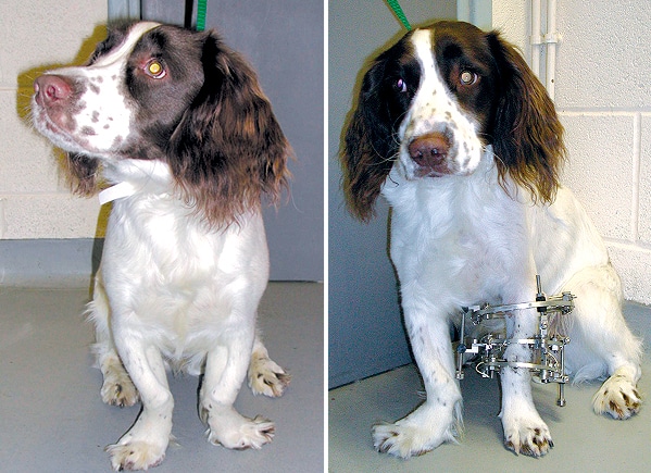
This Springer Spaniel has deformity of both fore limbs. The limbs are S-shaped with the paws deviating to the outside. The left deformity has been corrected and the cut bones stabilised with a circular external skeletal fixator.
Case study 3
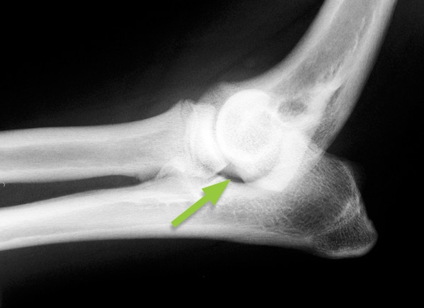
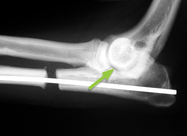
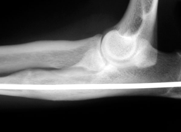
X-rays of a Cocker Spaniel with a painful elbow due to the two bones in the forearm, the radius and ulna, growing at different rates. The joint is deformed with a gap visible (small arrow). The ulna bone has been cut and lengthened so that the gap in the joint is no longer visible (large arrow). The bone has been stabilised with a metal pin. It healed within four months.
Case study 4
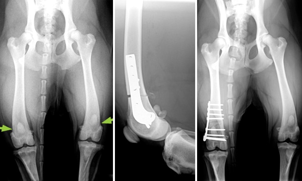
This Golden Retriever has deformity of both hind limbs with a resulting ‘bow-legged’ appearance (arrows). The more severely deformed femur (thigh bone) has been cut, straightened and stabilised with a special curved plate.
Case study 5
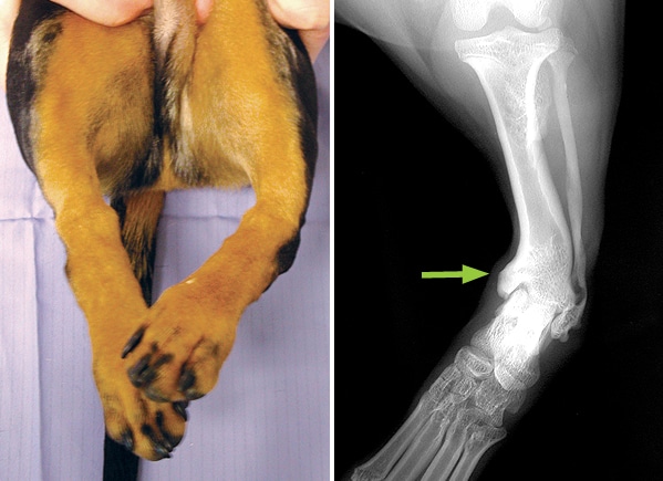
Photograph and X-ray of a Dachshund with severe deformity of the left hind limb. The tibia (shin bone) is bent just above the hock (ankle joint) (arrow) and the paw deviates inwardly.
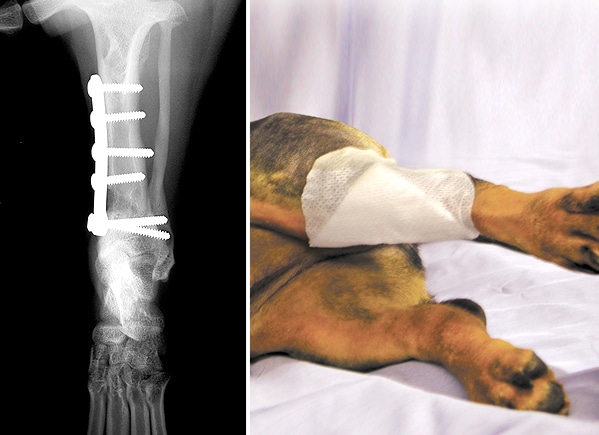
X-ray and photograph following surgery showing how the left tibia has been straightened and stabilised with a plate and screws.
Arranging a referral for your pet
If you would like to refer your pet to see one of our Specialists please visit our Arranging a Referral page.